Carrello: 0 Prodotto Prodotti (vuoto)

Nessun prodotto

Spedizione Non ancora calcolato
Totale: 0,00 €

Attributi:
Quantità:
Totale:

Ci sono 0 prodotti nel tuo carrello C'è 1 prodotto nel tuo carrello

Totale prodotti
Spese di spedizione  Gratuito!
Totale
Continua con lo shopping Vai al carrello
Prodotto aggiunto con successo alla carrello
Libri in lingua inglese

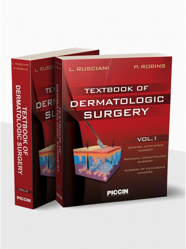
Textbook of Dermatologic Surgery

Autori
Rusciani - Robins

379,00 €

  • publish date agosto 2008
  • ISBN 978-88-299-1898-0
  • Code Piccin 1826701/2
  • Pages 960
  • Binding Cartonato
  • Accesso contenuti 3 anni dall'acquisto

Preface

The dynamic nature of the medical sciences presents a constant challenge for the dermatologist, but the past 25

years have necessitated a shift in approach to the profession. With the increasing influx of biological data, and developments in diagnostic and treatment techniques, our armamentarium has expanded to treat an increasing number of pathologies and enabled the vast advancement in corrective procedures. In addition, the recent prevalence of tumor pathologies––in particular, the tumors of the skin––has resulted in a growing number of patients to seek out the dermatologist for treatment.

Dermatologists are now required to continually update their knowledge of and techniques in dermatological surgery. As many as 350 pathologies can now be treated by surgery. Also dermatologists are now dealing with an increasing number of cosmetic surgery treatments, which require the use of several devices (eg, lasers, ultrasounds, and radiofrequencies, etc.). Such high-tech devices, and their evident biological action, can only be properly used by dermatologists after having acquired the proper expertise. Many countries all over the world have developed communities: national societies, conferences, and educational outlets, to address these surgical topics. This textbook was written in the spirit of these dermatological communities;

as a tool to improve surgical knowledge and technique; and, mostly, to assist the dermatologist in meeting the rising medical needs of today’s patient.

Luigi Rusciani

Perry Robins

Orders from Germany, Austria and Switzerland: www.schattauer.de

VOLUME I
GENERAL CUTANEOUS SURGERY
CHAPTER
1
THE DERMATOSURGICAL PATIENT
(G. Agostinacchio)
3
Introduction
3
Preparation of a Dermatologic Surgical
Procedure
3
Patient History
4
Preparation of a Healthy Patient for
Surgical Procedures
5
References
10
CHAPTER
2
INFORMED CONSENT IN CUTANEOUS SURGERY
(D.J. Goldberg)
11
New York § 2805-d
15
Iowa 147.37 Consent in Writing
15
Nebraska § 44-281
16
New Jersey title 8, §43G-4.1 Patient
rights
16
Connecticut §19-13 D3
16
CHAPTER
3
CUTANEOUS SURGERY IN OUTPATIENT AND OPERATING ROOM SETTINGS
(M. Simonacci, M. Sigona, A. Bettacchi,
R. Gasco)
19
The Dermatological Outpatient Surgery
Unit
20
The Operating Room
21
References
23
CHAPTER
4
SURGICAL INSTRUMENTS
25
(G. Guerriero)
25
Scalpels
25
Scissors
26
Needle Holders
26
Forceps
27
Hemostats
27
Curettes
28
Retractors
28
Towel Clamps
28
Containers and Forceps for Preparing
the Operative Field
28
Electrocoagulator
28
Dermabrader
29
Liposuction Apparatus
29
Miscellaneous Equipment and Supplies
29
Conclusions
29
CHAPTER
5
STERILIZATION AND DISINFECTION OF SURGICAL INSTRUMENTS
(M. Dal Canton)
31
Introduction
31
Definitions
31
Decontamination
31
Cleaning
32
Disinfection
32
Sterilization
32
Antisepsis
33
Decontamination
33
Cleaning
34
Sterilization
35
General Concepts
35
CONTENTS xiii
Heat Sterilization
37
Dry Sterilization
37
Steam Sterilization
38
Gas Sterilization
42
Chemical Disinfection
44
Glutaraldehyde
45
Ortho-Phtalaldehyde
46
Peracetic Acid
46
TAED (Tetra-Acetyl Ethylen-Diammine)
47
Instrument Care
48
Inspection and Care
48
Lubrication
48
Packaging of Surgical Instruments
49
Loading the Steam Autoclave
50
Testing and Validation of the Sterilization
Process
52
Physical Monitors
52
Chemical Monitors
53
Biologic Indicators
54
Issues on Dermatologic Surgery Instruments
55
Electrosurgical Units
55
Dermabrasion Equipment
55
Hair Transplant Equipment
56
Current Issues in Disinfection and
Sterilization of Surgical Instruments ..
56
References
57
CHAPTER
6
LOCAL ANESTHETIC TECHNIQUES
(R. Bono, G. Rossi)
61
Introduction
61
The Physiology of Pain Perception
61
Drugs Used to Produce Local and Regional
Anesthesia
62
The Mechanism of Action of Local
Anesthetics
62
The Chemical Structure of Local
Anesthetics
62
Vasoconstrictors
63
Adverse Effects of Local and Regional
Anesthesia
64
Local Adverse Effects
64p
Systemic Adverse Effects
64
Drug Interactions
66
Pregnancy
67
Preventing and Managing Side Effects ..
67
Pre-Anesthetic Evaluation of Patients
68
Anesthetic Techniques
69
Local Anesthesia
69
Regional Anesthesia (Peripheral Nerve
Block)
69
Sedation
70
Setting Equipment and Assistance
71
Patient Evaluation
71
Drugs used
71
References
72
CHAPTER
7
HAEMOSTASIS
(N. Frasca)
73
Physical Agents
73
Physiologic Agents
73
Topical-Chemical Agents
74
Thermal Devices
74
References
74
CHAPTER
8
SUTURING TECHNIQUES AND MATERIALS
(A. Cimitan)
75
Suturing Materials
75
Threads
75
Staples
77
Wound Closure Adhesive Tapes
78
Surgical Needles
78
Suturing Techniques
78
Most Common Suturing Techniques
79
Special Sutures
82
References
84
CHAPTER
9
TOPOGRAPHICAL ANATOMY AND CUTANEOUS SURGERY
(G. Leigheb, P. Boggio, G. Cristiani)
85
The Fronto-Occipital Region
85
The Temporal Region
86
The Auriculo-Mastoid Region
87
The Orbital Region
88
The Eyelids
88
The Lacrimal Apparatus
88
Vessels and Nerves
89
The Nasal Region
91
Nasal Support System
91
Superficial Structures
91
The Superficial Plains
91
Vessels and Nerves
93
The Lips
93
The Cheeks
94
The Hand
94
The Volar Surface of the Hand
94
The Dorsal Surface of the Hand
95
The Fingers
95
References
96
xiv
CHAPTER
10
CUTANEOUS BIOPSY
CONTENTS
Clinical Examples
117
References
120
(G. Guerriero)
97
Preparing the Patient
97
Planning the Biopsy
97
Disinfection and Anesthesia
98
Instruments
98
Biopsy Techniques
99
Incisional and Excisional Biopsies
99
Shave Biopsy
99
Punch Biopsy
99
Fine-Needle Biopsy
100
Nail Biopsy
100
Biopsy of the Oral Cavity
100
Complications
101
References
101
CHAPTER
11
CUTANEOUS FLAPS
I - Advancement Flaps
103
(J. Ocaña Sierra, M.L. Wilhelmi)
103
Basic Techniques
103
Rectangular Advancement Flap
104
Triangular Advancement Flap
104
Trapezoidal Advancement Flap
104
VY or YV Advancement Flaps
107
A-T Flap
107
Double Crescent Perialar Excision
107
Bipedicle Double Flap
108
Crescentic Advancement Flap
108
Special Techniques
110
Advancement Flaps of Suprauricular
Region
110
Advancement Flaps of Eyelids
110
Advancement Flaps of the Nose
110
Advancement Flaps of Lips
110
Advancement Flaps of the Cheek
and Chin
110
References
113
II - Rotation Flaps
114
(P. Robins, C.H. Schmults)
114
Classic Rotation Flap
114
Variations of the Rotation Flap
115
Hatchet Flap
115
Double Rotation Flaps
116
Techniques to Avoid Common Errors
117
Raise the Leading Edge
117
Widen the Tip of the Flap
117
Pay Attention to Cosmetic Units and
Skin Tension Lines
117
Consider the Effect of Secondary Motion
117
III - Transposition Flaps
121
(A. Rusciani, G. Guerriero, A. Paradisi)
121
Rhombic Flap
122
Classic Limberg Rhombic Flap
122
Dufourmentel Flap
123
Webster Flap
123
Rhombic Flap on a Circular Defect
124
Nasolabial Flap
126
Bilobed Flap
128
Z-Plasty
130
Complications
130
References
131
CHAPTER
12
CUTANEOUS GRAFTS
(A. Picoto)
133
Introduction
133
Types of Skin Grafts
133
Autografts
134
Cutting the Split-Thickness Skin Graft ..
135
Cutting the Full-Thickness Skin Graft ..
135
Donor Areas
135
Cutting the Graft
136
Mesh Grafts
138
References
138
CHAPTER
13
WOUNDS: CLASSIFICATION AND SURGERY
(R. Baiocchi, M. Serrai, G. Maccanti)
139
Introduction
139
Trophic Ulcers of the Lower Limbs
140
Venous Ulcers
140
Arterial Ulcers
141
Ulcers Due to Vasculitis
142
Infection-Based Ulcerations
142
Gangrene
145
Pressure Sores
146
Preparation for Surgery
147
Operative Technique
148
Review of Operative Techniques
148
Treatment of Multiple Pressure Sores
153
Conclusion
153
Diabetic Foot
155
Surgical Treatment
156
Conclusion
158
Traumatic Wounds
161
Contusion
162
CONTENTS xv
Excoriations
162
Lacerated and Contused Wounds
162
Flap Wounds
162
Defect Wounds
162
Pilonidal Sinus
166
Considerations and Conclusions
166
References
167
CHAPTER
14
SURGICAL WOUND DRESSING
(L.M. Chinni, M.T. Viviano, M. Papi)
171
Introduction
171
Types of Cutaneous Wounds
171
Physiology of Wound Healing
172
Phases of Wound Healing
172
What is the Ideal Characteristic of a
Wound Dressing?
172
Occlusive and Semi-Occlusive Dressing ..
173
Polymer Films
174
Polymer Foams
174
Hydrocolloids
174
Hydrogels
175
Alginates
175
Miscellaneous
176
Adhesive Tapes and Bandages
176
Tactifiers
176
References
176
CHAPTER
15
SURGICAL COMPLICATIONS
(K. Nouri, H. Chen, V. Vejjabhinanta,
A.R. Patel)
179
Introduction
179
Intra-Operative
179
Bleeding
179
Damage to Nerves or Surrounding
Vital Organs
180
Anesthetic Side Effects/Drug Interactions
181
Electrocautery Associated Complications
181
Post-Operative
181
Contact Dermatitis
181
Infection
182
Chondritis
182
Wound Dehiscence
183
Necrosis of the Skin
183
Suture Spitting
183
Suture Tracking
183
Excessive Granulation Tissue
184
Non-Healing Wounds
184
Pigment Change
184
Scars/Hypertrophic Scarring/Keloids
184
Conclusion
184
References
185
CHAPTER
16
PHOTOGRAPHIC DOCUMENTATION IN DERMATOLOGY
(E. Vigl, Pf. Zampieri)
187
Introduction
187
Rules for Correct Documentation
188
Try to Show Only the Clinical Sign
188
Pay attention to the Background
190
Choose the Right Detail
192
When Taking a Photograph Consider
the Angle Carefully
194
Using the Correct Lighting
194
Keep the Slides Clean
199
Do Not Make Inappropriate Use of
Other People’s Documentation
200
Do Not “Modify” Therapeutic Results
Obtained
200
Pay Careful Attention to the Order of the
Caption and the Written Text
200
Do Not Present An Excessive Number of
Slides
201
Respect the Privacy Data Protection Law!
201
References
202
REGIONAL DERMATOLOGIC SURGERY
CHAPTER
17
SURGERY OF THE EYELID REGION
(F. Camacho-Martínez)
205
Introduction
205
General Considerations
206
Anatomic Considerations
206
Lacrimal Apparatus
206
Functional Considerations
207
Surgical Considerations
207
Tarsorrhaphy
207
Surgical Techniques
208
Defects on the Lower Lids
208
Small Defects
208
Medium-Sized Defects
208
Complete Loss of the Lower Eyelid
213
Defects on the Upper Lids
217
Small Defects
217
Complete Loss of the Upper Lid
220
Skin Defects
221
Defects on the Lateral Canthus
224
Defects Only on the Lateral Canthus
224
Defects on the Lateral Canthus and
Small Defects of Whole Thickness on
Both Lids
225
Defects on the Lateral Canthus and Small
or Medium-Sized Cutaneous Defects on
Both Lids
225
Defects on the Inner Canthus
226
xvi
Defects on the Inner Canthus and
Small Defects of Whole Thickness on
CONTENTS
CHAPTER
20
SURGERY OF THE LIPS AND THE
Both Lids
226
Defects on the Inner Canthus and Small
or Medium-Sized Cutaneous Defects on
Both Lids
227
References
230
CHAPTER
18
AESTHETIC BLEPHAROPLASTY
(J. Carruthers, A. Carruthers)
233
The Presurgical Evaluation
233
History of Any Previous Peri-Ocular
Treatments
233
Evaluation of Subject’s Preoperative
Expectations
234
Necessary Documentation to Achieve
Surgical Goals
234
Cosmetic Upper Eyelid Blepharoplasty
237
Cosmetic Lower Eyelid Blepharoplasty
238
Traditional Infralash Blepharoplasty
238
Lateral Canthopexy
238
Skin Redraping
238
Transconjunctival Lower Eyelid
Blepharoplasty
238
Transconjunctival Blepharoplasty with
Anterior Fat Transposition: the Newest
Lower Eyelid Rejuvenative Procedure
239
Blepharoplasty Plus Resurfacing
239
Keratoconjunctivits Sicca
240
Complications and their Management
240
Complications Related to the Use of the
CO2 Laser for Aesthetic Blepharoplasty
241
Postoperative Care Regime
241
Ice Positioning and Wound Care
241
Summary
242
References
242
CHAPTER
19
NASAL PYRAMID SURGERY
(C. Alfano, A. Rusciani, S. Chiummariello,
C. Pagliarello)
243
Introduction
243
General Principles
243
Reconsctruction of the upper Part of the
Nose (Dorsum and Lateral Faces)
244
Reconstruction of the Lower Nose
245
Axial Flaps
247
Technical Tips
248
References
249
ORAL CAVITY
(W.A. Pagani)
251
General Considerations
251
I- Anatomy and Physiology
251
The Lips and Oral Cavity
251
Muscles
251
Innervation and Blood Circulation of the
Lips
252
Regional Lymphatic System
252
II- Cancer Ablative Surgery
252
Reconstructive Surgery
254
Lips: Superficial Defect Repair
254
Lips: Full-Thickness Defect Repair
255
Corner of the Mouth Reconstruction
257
Vermilion Border Restoration and
Principles of Oral Mucosa Surgery
258
Acknowledgments
265
References
265
CHAPTER
21
SURGERY OF THE AURICULAR REGION
(H. Breuninger)
267
Introduction
267
Ensuring Tumor-free Three Dimensional
Incision Margins (3D-Histology)
267
Indications
269
Pre-Therapeutic Diagnostics
270
Anesthesia
270
Planning the Operation
271
Operation
271
Suture Techniques
275
Aftercare
275
References
275
CHAPTER
22
SURGERY OF THE EXTREMITIES
(S. Serrano-Ortega)
277
Upper Extremities
278
Axillae
278
Arms and Forearms
280
Hands
280
Lower Extremities
283
Inguino-Crural Region
283
Thighs
283
Leg
285
Feet
289
References
291
CHAPTER
23
NAIL SURGERY
CONTENTS xvii
CHAPTER
26
SQUAMOUS CELL CARCINOMA
(E. Haneke)
293
Surgical Anatomy and Physiology of the
Nail
293
Preoperative Examination and Preparation
for Surgery
295
Anaesthesia and Instrumentation
296
Nail Avulsion and Biopsies
297
Characteristic Nail Lesions
299
Surgery of Benign Tumours
304
Longitudinal Melanonychia
305
Surgery of Malignant Tumours
306
Traumatic Lesions
308
Grafts and Flaps
308
References
309
SURGERY OF CUTANEOUS CANCERS
CHAPTER
24
BENIGN EPITHELIAL CANCERS
(G. Zumiani, L. Castellani)
315
Neoplasms of Viral Origin
315
Common Warts
316
Flat Warts
317
Condylomata Acuminata
317
Molluscum Contagiosum
318
Fibroepithelial Polyps
319
Seborrheic Keratosis
319
Dermatosis Papulosa Nigra
320
Stucco Keratosis
320
Clear-Cell Acanthoma
321
Keratoacanthoma
321
Comedone (Blackhead)
322
Verrucous Epidermal Naevus
322
Actinic Keratoses
323
Becker’s Naevus
324
Adnexal Neoplasm
325
References
334
CHAPTER
25
SURGICAL MANAGEMENT OF BASAL CELL CARCINOMAS
(J.F. Sei)
335
Tumor Size
337
Location
338
Histologic Type
338
Primary or Recurrent Tumor
340
Severity score
341
Results of Treatment
344
Conclusion
344
References
344
(J. Hafner, M. Hess Schmid, S. Läuchli,
L. Schärer, G. Burg)
347
Treatment Modalities and Prognosis of
Squamous Cell Carcinoma of the Skin
347
Precursors of Squamous Cell Carcinoma ..
351
Specific Variants of Low-Grade Squamous
Cell Carcinoma
355
Surgical Repair According to the Anatomical
Regions of the Head
357
Nose
358
Flaps and Grafts of the Nose
358
Ear
363
Flaps and Grafts of the Ear
363
Lips
364
Flaps and Grafts of the Lips
364
Cheek
366
Flaps and Grafts of the Cheek
367
Forehead
369
Flaps and Grafts of the Forehead
369
Scalp
371
Flaps and Grafts of the Scalp
371
References
373
CHAPTER
27
PIGMENTED LESIONS
(J.S. Conejo-Mir, A. Serrano,
L. Rodriguez-Freire, C. Hernandez)
377
Solar Lentigo
377
Treatment
377
Becker’s nevus
378
Clinical manifestation
378
Histologic Findings
379
Treatment
379
Congenital Nevi
379
Clinical Manifestations
379
Histological Features
380
Treatment
380
Acquired Nevocytic Nevi
384
Clinical and histological features
384
Management
384
Dysplastic Nevi
385
Clinical Features
385
Histological Features
385
Management
386
References
387
CHAPTER
28
MALIGNANT MELANOMA
(M.I. Perez, T.C. Yu)
391
References
395
xviii
CHAPTER
29
THE SENTINEL LYMPH NODE
CONTENTS
Diagnosis of Pigmented Skin Lesions
426
Differentiation Between Melanocytic
(G. Landi, M. Polverelli, C. Landi)
397
Introduction
397
The Orderly Lymphatic Progression of
Melanoma
398
Morton’s Experience
400
Preoperative Lymphoscintigraphy
401
Intraoperative Gamma Probe
404
Surgical Technique
406
Histopathologic Investigation
408
Regional Complete Nodal Dissection
410
New Strategies
411
Sentinel Node Biopsy in Other Tumors
412
Personal Results
412
Complications
414
Limits of the Procedure and Controversies
414
References
416
CHAPTER
30
EPILUMINESCENCE MICROSCOPY
(R. Pellicano, M. Lomuto)
419
Introduction
419
Historical Overview
419
Observation Techniques
420
Dermoscopic Criteria
420
Pigment Network (Typical Atypical)
421
Streaks (Regular Irregular)
421
Dots/Globules (Regular Irregular)
422
Blue-Whitish Veil
423
Regression Structures (White Areas/Blue
Areas)
423
Hypopigmented Areas (Focal Multi-Focal
Diffuse)
423
Blotches (Localised Diffuse Regular
Irregular)
423
Multiple Milia-Like Cysts
424
Comedo-Like Openings
Leaf-Like Areas
424
Red-Blue Lacunas
424
Vascular Pattern
424
and Non-Melanocytic Lesions
426
Differentiation Between Melanocytic
lesions and melanoma
427
Conclusions
431
References
432
CHAPTER
31
MALIGNANT TUMORS OF THE DERMIS AND SUBCUTANEOUS TISSUE
(P.G. Lang, Jr.)
435
Merkel Cell Carcinoma
435
Malignant Granular Cell Tumor
437
Pilomatrix Carcinoma
438
Sebaceous Gland Carcinoma
438
Sarcomas
440
Dermatofibrosarcoma Protuberans
440
Atypical Fibroxanthoma
442
Malignant Fibrous Histiocytoma
443
Leiomyosarcoma
444
Angiosarcoma
446
Epitelioid Sarcoma
448
Fibrosarcoma
449
Liposarcoma
450
Cutaneous Rhabdomyosarcoma
450
Malignant Peripheral Nerve Sheath
Tumor
450
Sweat Gland Carcinomas
450
Aggressive Digital Papillary Adenoma ..
451
Signet Ring Carcinoma
451
Malignant Eccrine Spiradenoma and
Malignant Cylindroma
451
Mucinous Carcinoma
452
Malignant Mixed Tumor
452
Malignant Clear Cell Hidradenoma
452
Porocarcinoma
453
Primary Cutaneous Adenoid Cystic
Carcinoma
454
Apocrine Carcinoma
454
Microcystic Adnexal Carcinoma
455
References
457
CONTENTS xix
VOLUME II
SPECIAL CUTANEOUS SURGERY
CHAPTER
32
CURETTAGE AND ELECTRODESICCATION
(Arash Kimyai-Asadi, Ming H. Jih)
473
Introduction
473
Procedure
473
Efficacy
474
Indications and Contraindications
475
Conclusions
475
References
476
CHAPTER
33
CRYOSURGERY
(P. Mintzias)
477
Historical Background
477
Cryobiology
477
Vascular changes
479
Cryoimmunology
479
Equipment - Techniques of treatment
479
Dipstick Technique
480
Cryoprobe Technique
481
Spray Technique
482
Intralesional Technique (Weshahy’s
Technique)
483
Freezing Parameters
483
Monitoring Method
484
Use of Thermocouple Measurements
484
Impedance-Resistance Methods
485
Indications and Contraindications
485
Cryosurgery in Benign Skin Tumours
486
Seborrhoeic Warts
486
Haemangiomas
487
Keloids and Hypertrophic scars
487
Pyogenic Granuloma
487
Warts
488
Cryosurgery in Skin Precancerous
Lesions
490
Actinic Keratosis
490
Bowen’s Disease
490
Queyrat’s Erythroplasia
491
Leucoplakia
491
Lentigo Maligna
492
Keratoacanthoma
492
Cryosurgery in Skin Cancer
492
Basal Cell Carcinoma (BCC)
493
Squamous Cell Carcinoma (SCC)
497
Malignant melanoma
498
Complications of Cryosurgery
499
Perspectives – Conclusion
499
References
500
CHAPTER
34
ELECTROSURGERY
(E. Sbano, P. Sbano)
505
History
505
Basic Principles
506
Electrical Circuits
506
Waveforms
506
Electrodes
506
Devices
508
Techniques
508
Clinical Applications
509
Hazards
514
Comparison of Electrosurgery with other
Surgical Methods
516
References
517
CHAPTER
35
DERMABRASION
(J.M. Yarborough, C.B. Harmon)
519
Introduction
519
Indications
519
Preoperative Consultation and
Examination
520
Preoperative Medications and Laboratory
Evaluation
520
Surgical Technique
521
Postoperative Wound Care
523
Complications
524
References
525
CHAPTER
36
TISSUE EXPANDERS
(I. Zilinsky, Y. Har-Shai, G. Weiss)
527
Microstructure of the Human Skin
527
Terminology
527
Bio-Mechanical Properties of Skin
528
Historical Precedence of Stretching Skin ..
530
Histological Changes
531
Epidermis
531
Dermis
531
Adnexa
532
Subcutaneous Fat
532
Muscle
532
Vascularity
532
Capsule
532
Long-term Histopathological Results
533
Linear Tissue Expansion
533
Types of Linear Skin-Stretching Devices
534
The sure-closure® skin-stretching system
xx CONTENTS
(SSD) (Zimmer)
534
The Suture Tension Adjustment Reel
(STAR)
536
The External Tissue Extender (ETE® –
Höjmed AB Sweden)
536
The Frechet Scalp Extender
536
The Constant-Tension Approximator
(Proxiderm – Progressive Surgical Products
Westbury New York)
536
Spherical Tissue Expansion
539
Clinical Applications of Spherical Devices
539
Breast Reconstruction with a Spherical
Expander
540
Giant Congenital Nevi
541
Complications
542
References
543
CHAPTER
37
SCAR REVISION
(A. Tulli, A. Diociaiuti)
547
Introduction
547
Indications
547
Types of Scars
548
Hypertrophic Scars
548
Keloids
548
Depressed Scars
548
Widened Scars
548
Long Linear Scars
548
Pitted Scars
548
Trapdoored Scars
548
Contracted Scars and Webbed Scars
549
Distortion of Free Margins
549
Red Scars
549
Surgical Technique
549
Fusiform Excision
549
Skin Flaps
549
Dermal Micrografts
550
Autologous Grafts
551
Dermoabrasive Scar Revision
551
Laser Scar Revision
552
Non-Surgical Scar Revision
552
Corticosteroids
552
Pressure
552
Radiation Therapy
552
Cryotherapy
552
Medical Therapy
552
References
553
CHAPTER
38
TREATMENT OF VARICOSE VEINS
(S. T’ Kint, D. Roseeuw)
555
Introduction
555
Anatomy
555
Venous Pathology
556
Negative Factors
556
Positive Factors
556
Clinical Features
556
The Schwartz Test (Wave Test)
557
Cough Test
557
Trendelenburg test
557
Perthes Test
558
Pratt Test
558
Linton Test
558
Duplex Ultrasonography
558
Ankle-Arm Index
558
Venography
558
Air Pletysmography
559
Treatment
559
Drugs
559
Mechanical therapy
559
Sclerotherapy
560
Ambulatory Phlebectomy
561
References
564
CHAPTER
39
SCLEROTHERAPY
(S. Rapprich, E. Hasche, M. Hagedorn)
565
Introduction
565
Varicosis
565
Definition of Varicosis
565
Epidemiology
565
Etiology
565
Pathophysiology of Varicosis
566
Clinical investigation and machine
diagnostics
566
Phlebologic Anamnesis
566
Clinical Picture
567
Phlebological Investigation
568
Chronic Venous Insufficiency (CVI)
571
Skin Changes with CVI
572
Therapy of Varicosis
577
History of Sclerotherapy
578
Indications of Sclerotherapy
579
Therapeutic Basics
580
Sclerosing Agent
580
Pathophysiology of Sclerotherapy
581
Practical Execution
582
Puncture and Air-Block Technique
582
Tournay Sclerosing Method
582
Injection
583
Sigg Sclerosing Method
583
Sclerosing Intradermal Varicose Veins ..
584
Therapeutic Procedure
585
Supplementary Measures
585
CONTENTS xxi
Contraindications
586
Complications
586
Topical Complications
586
Systemic Reactions
589
Follow-up Visits
590
References
590
CHAPTER
40
LASERS AND POLYCHROMATIC LIGHT SOURCES (PCLS) IN DERMATOLOGY
(L. Marini)
591
Light-Tissue Interaction
591
Light-Sources
593
Lasers
593
PCLS (Polychromatic Light Sources)
594
Dosimetry
595
Cooling Systems
595
Surgical Lasers
598
Vascular Targets - Vascular Specific Lasers
and IPLS
601
Pigmented Targets: Pigment-Specific
Lasers and IPLS
606
Non-Ablative Collagen Remodeling and
Skin Rejuvenation
609
Conclusion
611
References
611
CHAPTER
41
THE CARBON DIOXIDE LASER
(M.A. Trelles, R.G. Calderhead)
615
History and Photobiosurgical Basics
615
Photothermal Bioreactions
615
Irradiance (Power Density) – The Surgical
Key
618
Radiant Flux (Energy Density) – the Laser
“Dose”
620
Beam Mode
621
Beam Type
621
Beam Transmission
623
Illustrative Examples
624
Incision
624
Excision
624
Vaporization
625
Coagulation
626
Vaporization and Coagulation Combined
626
Variations on a Theme (CO2 ‘Pinhole’ Treatment)
626
CO2 Laser-Assisted Blepharoplasty
628
CO2 Laser: Quo Vadis?
629
CHAPTER
42
LASER SAFETY WITH THE CO2 LASER
(M.A. Trelles, R.G. Calderhead)
631
CO2 Laser-Related Hazards
631
Electrical Hazards
631
Skin or Other Tissue Hazards
632
Carelessness
632
Misalignment of the Aiming Beam
632
Accidental Irradiation from Beam
Reflection
632
Tissue Protection for the CO2 Laser
632
Optical Hazards
633
Fire Hazards
634
Laser Plume Hazard
634
Side Effects
634
Common Sense and Hazard Prevention
635
CHAPTER
43
LASER THERAPY IN VASCULAR LESIONS
(J.M. Fernández Vozmediano,
J.C. Armario Hita)
637
Introduction
637
Treatment of Vascular Lesions
638
Telangiectasias
638
Hemangiomas
639
Vascular Malformations
642
Laser Therapy in Vascular Lesions
642
Continuous-Wave Systems
643
Argon Laser
643
Argon-Pumped Tunable Dye Laser
(APTDL)
645
Potassium Titanyl Phosphate
532
Nanometer Laser (KPT Laser)
645
Copper Vapor Laser
646
Krypton Laser
646
Copper Bromide Laser
646
Pulsed-Wave systems
647
Action on Tissues
647
Method of Application
647
Contraindications
648
Complications
648
Indications
649
Telangiectasias
649
Vascular Malformations
651
Hemangiomas
653
Other Vascular Lesions
654
Other Laser Systems Used in Vascular
Lesions
654
Nd:YAG laser
654
CO2 laser
655
Treatment of Telangiectasias of the Legs
656
Sclerotherapy
656
xxii
CONTENTS
Laser Therapy of Leg Veins
657
Dye Laser Systems
657
Nd:YAG Systems
658
Long-Pulsed Alexandrite Laser
659
Diode Laser
659
Systems of Non-Coherent Light
659
References
660
CHAPTER
44
ERBIUM LASERS
(M.G. Onesti)
665
Characteristics
665
Applications
666
Erbium Laser and Resurfacing
666
References
667
CHAPTER
45
HISTORY OF MOHS MICROGRAPHIC SURGERY
(C.W. Hanke, A.L. Leonard)
669
The Early Years: the Fixed-Tissue
Technique
669
Modern Mohs Micrographic Surgery: the
Fresh-Tissue Technnique
671
Early Pioneers in Mohs Surgery
671
Modern Mohs Micrographic Surgery:
Expanding Horizons
673
The Legacy of Frederic Mohs
674
References
674
CHAPTER
46
MOHS MICROGRAPHIC SURGERY: A TREATMENT OF FIRST CHOICE FOR DIFFICULT SKIN CANCERS
(N.W.J. Kelleners-Smeets, H.A.M. Neumann)
677
Introduction
677
History
678
Method
679
Indications for Mohs Micrographic
Surgery
682
Basal Cell Carcinoma
682
Squamous Cell Carcinoma
684
Discussion
684
Conclusion
688
References
688
COSMETIC SURGERY
CHAPTER
47
CUTANEOUS AGING: ALTERED SKIN TROPHISM AND WRINKLES
(M.T. Leccia, X. Martinet)
693
Intrinsic Aging and Photoaging: Clinical
and Functional Aspects
693
Intrinsic Aging and Photoaging:
Histologic Features
694
Intrinsic Aging
694
Photoaging
695
Pathogenesis of Photoaging: Current
Concepts
696
Prevention of Cutaneous Aging
697
References
700
CHAPTER
48
CHEMICAL PEEL: CLASSIFICATION, INDICATIONS
(A.D. Katsambas, E. Nicolaidou)
703
Classifications of Peeling Agents
703
Indications of Chemical Peels
705
Facial Skin Peels
706
Glycolic Acid Peels
706
TCA Peels
707
Pesorcinol Peels
708
Phenol Peels
708
Salicylic Acid Peels
709
Non-Facial Skin Peels
709
References
710
CHAPTER
49
CHEMICAL PEELS
(Ming H. Jih, Arash Kimyai-Asadi)
711
Introduction
711
Procedure
712
Complications
713
References
713
CHAPTER
50
CHEMICAL PEELING WITH TCA
(A. Camps-Fresneda, G.A. Moreno-Arias)
715
Introduction
715
Chemical Characteristics of TCA
715
TCA Formulation
715
What Concentration?
715
Clinical and Histological Aspects
716
CONTENTS xxiii
Indications and Contraindications
717
Skin Preparation
718
The Day of the Peeling
718
Written Informed Consent & Photographs ..
719
Anesthesia and Sedation
719
Materials
719
Technique
719
Postoperative Care
722
Side Effects and Complications
722
Re-Peeling
723
Non-Facial Areas
723
TCA Combined with Other Agents
725
Conclusion
726
Acknowledgments
726
References
726
CHAPTER
51
GLYCOLIC ACID: INDICATIONS AND APPLICATION
(G. Vezzoni, G.M. Vezzoni)
727
How Superficial Peeling with Glycolic
Acid Works
727
History
728
What Peeling Does
728
What Peeling Does Not Do
728
Carrying Out Superficial Peeling with
Glycolic Acid
729
Preparing the Patient
729
Technique for Peeling the Face with 70% Glycolic Acid
729
Summary
731
References
731
CHAPTER
52
DEEP CHEMICAL PEELING
(G.D. Monheit)
733
The Peel
737
References
740
CHAPTER
53
DERMOFILLERS: COLLAGEN, HYALURONIC ACID, SILICONE, METHACRYLATE ETC
(D. Dasiou-Plakida)
741
Introduction
741
Fluid Silicone
741
Fluid Medical Grade Silicone
741
Tissue Histological and Immuno-
Histological Reaction
743
Injection Techniques
744
Methodology
745
Indications
746
Adverse Reactions
746
Animal Collagen Implants (Zyderm I
Zyderm II Zyplast)
747
Skin Test
747
Injection Techniques
748
Scars
748
Nasolabial Furrows and Wrinkles
749
Glabellar Wrinkles
749
Perioral Vertical Lines
749
Horizontal Forehead Wrinkles
749
Periorbital Wrinkles
749
Lip Augmentation
749
Results and Duration of Correction
750
Adverse reactions
750
Fibrel
750
Artecoll
751
Resoplast
753
Kokken Atelogen
753
Injectable Human Tissue Matrix-
Dermalogen
753
Isolagen
754
Autologen
754
e-Ptee (Soft Form)
755
Gore-Tex
756
Alloderm
756
Products of Hyaluronic Acid ...

High-Tech Dermo

Dimensione del file (17.08k)

Recensione JDD

Dimensione del file (17.25k)

No customer comments for the moment.

Scrivi la tua recensione

Prodotti correlati